Why Do Amphibians Have Moist Skin? Breathing and Water
Amphibian skin is moist because it does several jobs at once. Amphibians breathe through their skin as well as their lungs, so the skin must stay damp. It is also permeable, which means water passes in and out through it. Some species make mucus to keep it wet, and others, like toads, have drier-looking skin. Dry skin is a risk, and so is anything that disrupts skin function.
Amphibian skin is thin, scaleless and unprotected. That makes it a good breathing surface and a weak barrier to water loss.
This article explains why moisture matters, how amphibians manage water through the skin, and what goes wrong when the skin fails. For the breathing side, see how amphibians breathe in and out of water.
How this was made: prepared with AI assistance and checked against the sources listed at the end. Images are AI-generated illustrations, not photographs of real animals or products.
Key Takeaways
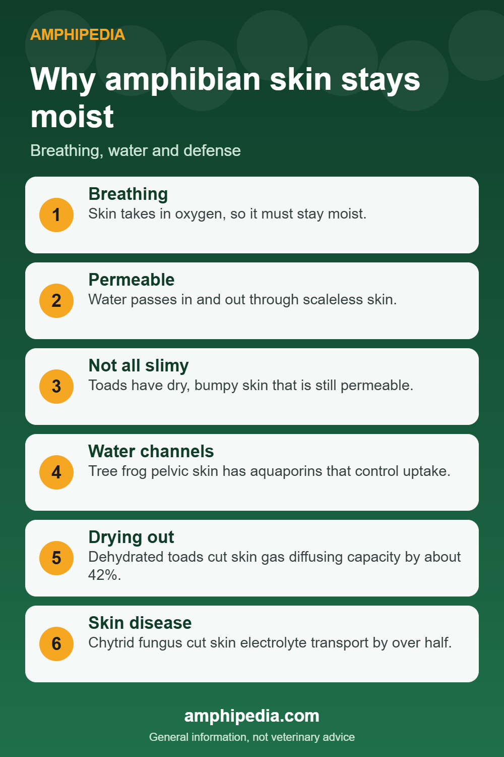
- Amphibians breathe through their skin, so it must stay moist.
- Skin is permeable to water, so amphibians can dry out.
- Not all amphibian skin is slimy; toads have dry, bumpy skin.
- Frog skin has water channels called aquaporins that control water uptake.
- A skin fungus can kill amphibians by upsetting salt balance.
Pin this infographicShare on Facebook
Skin that does several jobs
Amphibian skin is scaleless and permeable to water, according to the Pennsylvania Fish and Boat Commission. Adults usually breathe with lungs, but they can also absorb oxygen through the skin, a process called cutaneous respiration.
Lincoln Park Zoo makes the same point in simple terms. Amphibians breathe through both lungs and skin, so they must keep their skin moist and often live in humid places.
Skin traits and what they do
| Trait | What it does | Evidence |
|---|---|---|
| Moist surface | Allows gas exchange through the skin | PA Fish and Boat; Lincoln Park Zoo |
| Permeable skin | Lets water enter and leave | PA Fish and Boat; Burggren and Vitalis 2005 |
| Aquaporins | Water channels that raise water uptake | Journal of Experimental Biology study of a tree frog |
| Granular glands | Store toxins in some species | Smithsonian’s National Zoo |
| Dry, bumpy skin | Found in toads; still permeable | PA Fish and Boat; Britannica |
A leaky but managed barrier
A 2005 study in the Journal of Experimental Biology by Burggren and Vitalis looked at Woodhouse’s toad. It describes amphibian skin as a dynamic barrier to both water loss and uptake. The paper cites earlier work showing that toad skin offers substantial resistance to water loss compared with an open water surface.
When the toads were dehydrated, they became highly active, likely looking for water, then adopted postures that reduced their exposed surface. Rehydration was faster than dehydration in every test.
Frog skin also has dedicated water channels. A study in the same journal found that the ventral pelvic skin of the Japanese tree frog contains two aquaporins that control how much water the skin absorbs. Hormones such as arginine vasotocin moved these channels into position and raised water permeability.
Not all amphibians are slimy
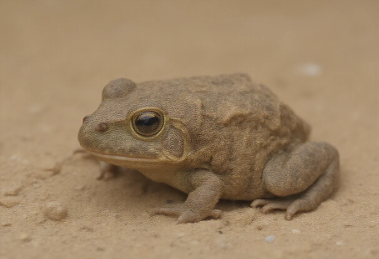
Some species have moist, slimy skin from mucus. Others, like toads, have dry, bumpy skin, the Pennsylvania commission notes. Britannica describes true toads as having thick, dry, warty skin.
Dry-looking skin does not mean impermeable skin. Toads still depend on skin and lung breathing, and the Burggren and Vitalis study was done on a toad.
Lungless salamanders
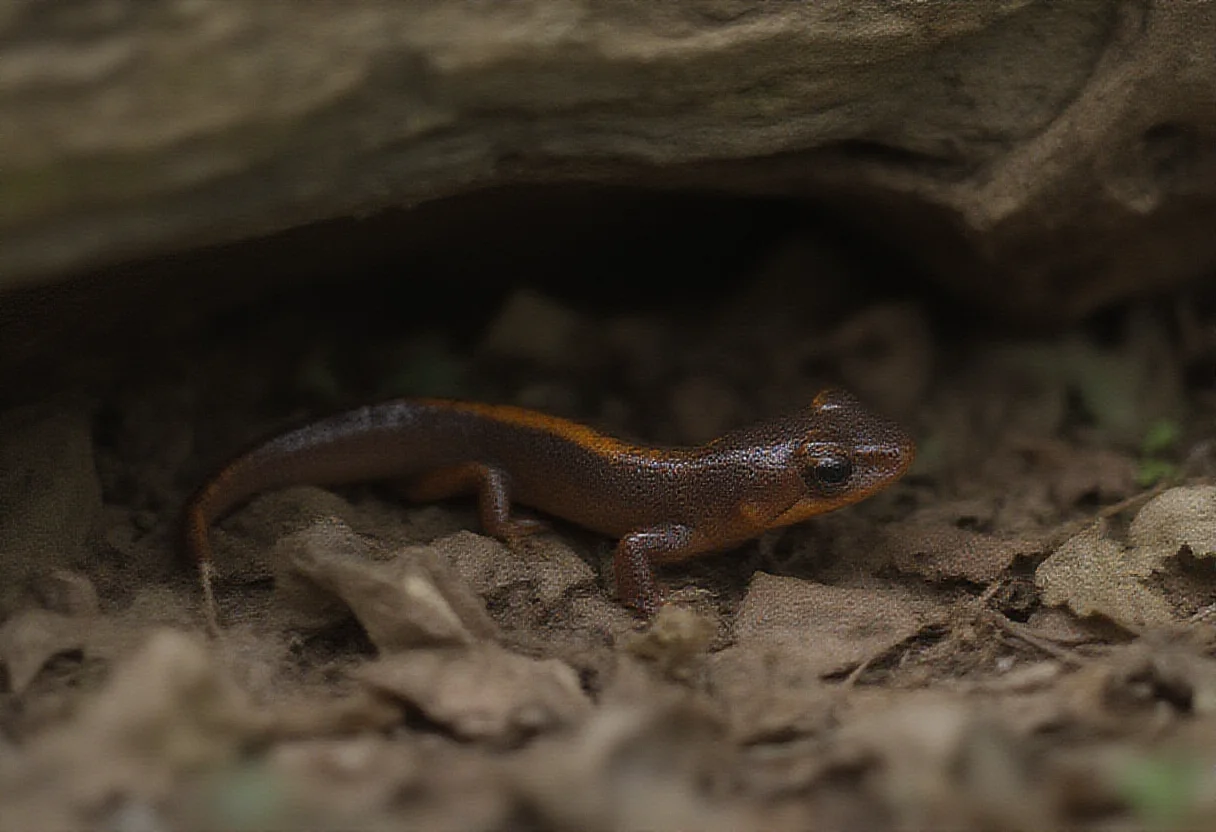
The clearest case of skin dependence is the lungless salamander family. A 2022 Harvard-led study in Science Advances reports that its adults have no lungs and breathe through the skin and the mucus membranes of the mouth and throat. The Smithsonian’s National Zoo says the eastern red-backed salamander must live in damp places to breathe. See whether salamanders have lungs.
When skin fails
Because skin controls fluid and salt balance, damage to it can be deadly. A 2009 Science study of chytridiomycosis, a disease caused by the fungus Batrachochytrium dendrobatidis, tested infected green tree frogs. Their skin’s ability to transport electrolytes fell by more than half, blood sodium and potassium dropped, and the frogs died of cardiac arrest.
The authors suggest that disrupted skin function may explain how the fungus kills many unrelated amphibian species. For the wider picture, see why amphibians are so vulnerable.
The seat patch: where a toad drinks
Toads rarely drink by mouth. A 2002 presentation by Viborg and Hillyard to the American Physiological Society, covered by ScienceDaily, studied the pelvic skin of Woodhouse’s toads and red-spotted toads, called the seat patch. It makes up about 10 percent of the skin area but accounts for over 70 percent of water uptake in dehydrated toads.
Blood flow in the patch rose seven to eightfold after water exposure, reaching near its peak within one minute. The toads adopted their water absorption posture during or shortly after that peak. This was a conference report on 11 toads, so treat it as a clear example, not a rule for every species.
Lungless salamanders: the extreme case, in more detail
Harvard’s Museum of Comparative Zoology reported in 2022 that lungless salamanders of the family Plethodontidae form lung-like structures as embryos, even though every adult lacks lungs. The structure disappears before hatching. Adults breathe through the skin and the lining of the mouth and throat.
The same report says lung loss has happened independently at least four times among amphibians, and that plethodontids make up more than two-thirds of living salamander species. The genetic pathways for lung development have been at least partly conserved despite at least 25 million years without functional adult lungs.
Chytrid and the cost of a failing skin
The 2009 Science study by Voyles and colleagues gives the numbers behind the chytrid discussion above. In infected green tree frogs, skin electrolyte transport fell by more than 50 percent, blood sodium fell by about 20 percent, and potassium by about 50 percent.
The cause of death was asystolic cardiac arrest, meaning the heart stopped beating. The authors suggest that because skin helps maintain internal balance, disrupted skin function could explain how the fungus kills many distantly related species.
Rollins-Smith and colleagues add that chytrid zoospores stay motile for about 24 hours at 23 C and survive more than 48 hours at 4 to 14 C. These are laboratory figures for the spore stage of the fungus, not a measure of risk in any particular pond.
Skin as a chemical defense
A 2011 review in Integrative and Comparative Biology by Rollins-Smith and colleagues explains that granular glands in the skin make antimicrobial peptides. The peptides are stored in granules and expelled onto the skin when nerve signals make cells around the gland contract. The gland then regenerates its stores.
Purified peptides inhibit chytrid growth in laboratory tests, working better against motile zoospores than mature spores. In Xenopus laevis, depleting peptides made frogs more susceptible to infection. Frogs release more peptide when chased, reaching levels that can inhibit the fungus.
The review adds limits. Not all amphibians make these peptides, and some families lack them. In some species the granular glands mature only at metamorphosis, which may leave newly metamorphosed frogs vulnerable. Stress hormones such as corticosterone can slow peptide renewal.
| Skin feature | Job | Where it is described |
|---|---|---|
| Moist surface | Gas exchange | PA Fish and Boat; Lincoln Park Zoo |
| Aquaporins | Water uptake | Journal of Experimental Biology |
| Seat patch | Most water uptake in toads | Viborg and Hillyard (2002) |
| Granular glands | Store peptides and toxins | Rollins-Smith et al. (2011); Smithsonian |
| Electrolyte transport | Salt balance | Science (2009) |
What thin, active skin means for keepers
This is general information, not veterinary advice. The Royal Veterinary College axolotl care sheet asks for minimal handling because the skin is vulnerable to toxins and trauma. It also advises a de-chlorinator for tap water, and calls one essential where chloramines are used.
Those points are about axolotls, so they are not a universal rule. Still, a reasonable reading of the skin biology above is that for any pet amphibian, water quality and gentle handling matter more than they do for scaled animals.
- Keep handling to a minimum.
- Treat water chemistry as part of the animal’s health.
- Photograph unusual skin changes and describe when they started.
- See an exotics vet from the ARAV directory rather than treating at home.
For people, the CDC says reptiles and amphibians often carry Salmonella. Wash hands with soap and running water after contact. Children younger than 5 years old should not handle or touch reptiles or amphibians or their environments.
Common mistakes about amphibian skin
- Assuming toads do not need water because their skin looks dry. The seat patch shows they take up a lot through it.
- Treating all amphibians as slimy. Toads have thick, dry, warty skin.
- Calling skin permeable and therefore weak. It is also a managed barrier and a chemical defense.
- Overlooking stress. Corticosterone can slow peptide renewal, according to Rollins-Smith and colleagues.
- Applying one species’ care rules to another. Handling and water needs differ.
Frequently Asked Questions
Why do amphibians need moist skin?
Because they absorb oxygen through the skin, and that works only while it stays moist.
Is amphibian skin waterproof?
No. It is scaleless and permeable to water, so amphibians can lose and take up water through it.
Why do toads have dry skin?
Toads have thick, dry, warty skin, but it is still permeable and they still need access to moisture.
Can amphibians drink through their skin?
Frogs take in water through their skin. A study of a tree frog found aquaporins in its pelvic skin that control water uptake.
What happens if amphibian skin dries out?
The animal can lose water faster than it can replace it. Dehydrated toads in one study became very active and changed posture to cut exposed surface.
Why is amphibian skin vulnerable to disease?
Skin controls salt and fluid balance. Chytrid fungus cut electrolyte transport in infected frog skin by more than half.
Bottom Line
Moist skin lets amphibians breathe and manage water, but it also ties them to damp habitats. Species such as toads have adjusted with drier skin and by changing posture when they dry out.
The same thin skin explains many risks, from dehydration to a deadly skin fungus.
Sources
- Pennsylvania Fish and Boat Commission: What is an amphibian?, cited for permeable skin, skin breathing and toad skin
- Lincoln Park Zoo: What is an amphibian?, cited for breathing through lungs and skin and for moist habitats
- Burggren and Vitalis (2005), Journal of Experimental Biology 208:105-112, cited for skin water loss and gas exchange in a toad
- Journal of Experimental Biology 213(2):288 – aquaporins in tree frog skin, cited for aquaporins in ventral pelvic skin
- Science (2009): Pathogenesis of chytridiomycosis, cited for chytrid disrupting skin electrolyte transport
- ScienceDaily: lungless salamanders (Science Advances, 2022), cited for skin breathing in Plethodontidae
- Smithsonian’s National Zoo: Eastern red-backed salamander, cited for lungless anatomy and the need for damp habitat
- Britannica: Toad, cited for dry, warty toad skin
- Smithsonian’s National Zoo: After an insect detox, can once-poisonous frogs get their spice back?, cited for granular glands under the skin
- ScienceDaily (2002): Toads and the seat patch, Viborg and Hillyard, American Physiological Society meeting, accessed 13 November 2026
- Rollins-Smith et al. (2011): Amphibian immune defenses against chytridiomycosis, Integrative and Comparative Biology, accessed 13 November 2026
- Royal Veterinary College Exotics Service: Axolotl care sheet (2024), accessed 13 November 2026
- CDC: Reptiles and amphibians and Salmonella, accessed 13 November 2026
- Harvard Museum of Comparative Zoology (2022): Lungless salamanders develop lungs as embryos despite lung loss in adults for millions of years, accessed 13 November 2026
- Voyles et al. (2009): Pathogenesis of chytridiomycosis, Science (abstract), accessed 13 November 2026